WHY AVOID SUTURES IN PTERYGIUM SURGERY
Uncomfortable pterygium surgery with stitches
Historically, pterygium surgery results markedly improved once conjunctival autografts started being used. Here a piece of healthy tissue is relocated from under the eyelid to the area of removed pterygium - assisting healing and reducing recurrence rates.
To facilitate this development, microscopic sutures were crucial to its success and longevity - preventing the grafted tissue from dislodging. However, they have always caused a significant degree of discomfort in the post-operative period, often for weeks.
Modern techniques have built upon the autograft innovation, using that design with a biological glue to avoid the patient discomfort that comes with stitches.
A glue revolution
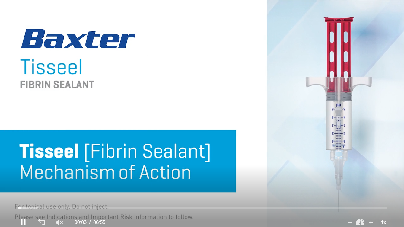
The development of biological tissue adhesives — most notably fibrin sealants such as Tisseel — represents one of the most quietly transformative advances in modern surgery. Fibrin glue mimics the final stage of the body's own clotting cascade, creating a natural, flexible bond between tissues without the need for sutures. Across surgical disciplines from cardiac surgery to neurosurgery, the ability to halt bleeding with a biological adhesive rather than mechanical stitches has reduced operative time, minimised inflammation, and improved healing. In ophthalmology, where the structures being operated on are measured in fractions of a millimetre and any source of post-operative irritation is magnified by the sensitivity of the ocular surface, the impact has been particularly significant. Nowhere is this more apparent than in pterygium surgery. The traditional sutured conjunctival autograft — while effective — left patients with fine stitches on the surface of the eye for weeks, driving inflammation, discomfort, and in some cases contributing to the very recurrence the surgery aimed to prevent.
The adoption of fibrin glue fixation fundamentally changed this. The graft adheres immediately, integrates naturally, and the eye is spared the prolonged foreign-body response that sutures inevitably produce. The result is faster healing, a more comfortable recovery, a cleaner cosmetic outcome, and — in experienced hands — recurrence rates that consistently sit below one percent. What was once considered a technically demanding alternative to suturing is increasingly recognised as the gold standard in pterygium surgery worldwide.
Why technique matters for recurrence
Pterygium recurrence rates vary dramatically by technique. Simple excision alone carries recurrence rates as high as 70–80% in some studies - a technique which has been thoroughly superseded. Conjunctival autograft performed by an experienced surgeon, reduces this to below 1%.
A recurring pterygium is harder to treat the second time. Scar tissue from the first surgery complicates the anatomy, limits graft options, and increases the risk of complications. Getting it right the first time is not optional.
There is no question that pterygium surgery needs a conjunctival autograft, the only decision is how to secure it to your eye - uncomfortable stitches or revolutionary glue
| Factor | No-stitch autograft (fibrin glue) | Bare sclera excision | Autograft with sutures |
|---|---|---|---|
| Recurrence rate | Very low Very low <1% in experienced hands |
Very high Up to 70–80% |
Low Very low <1% in experienced hands |
| Post-op comfort | Minimal irritation for 24–48 hours only | Moderate discomfort | Significant suture irritation for weeks |
| Healing speed | Faster graft adhesion | Slow | Sutures typically dissolve in 4–6 weeks |
| Cosmetic result | Excellent — natural-looking, minimal scarring | Poor — high recurrence, scarring | Good — may show suture tracks |
| Inflammation | Reduced — glue is biologically inert | Moderate to high | Moderate — sutures drive inflammation |
| Surgical complexity | High — demands precision and experience | Simple | Moderate |